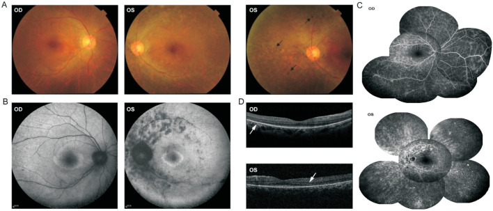
Figure 2. Retinal images and SD-OCT images of Family 2.
The fundus examination of the patient shows no abnormalities in the right eye and peripheral intraretinal bone spicule pigmentation (black arrow) in the left eye (A). The FAF image reveals an inhomogeneous hyperautofluorescent ring surrounding central hypoautofluorescence in each eye, and several anomalous scattered atrophic hypoautofluorescent scars along the supratemporal and infratemporal vascular arcade in the left eye (B). The fundus FFA images show remarkable hyperfluorescence at early stage in each eye (C). The SD-OCT scans show a loss of the outer retina outside of the central macula in each eye (white arrow; D). OD: Right eye; OS: Left eye.

