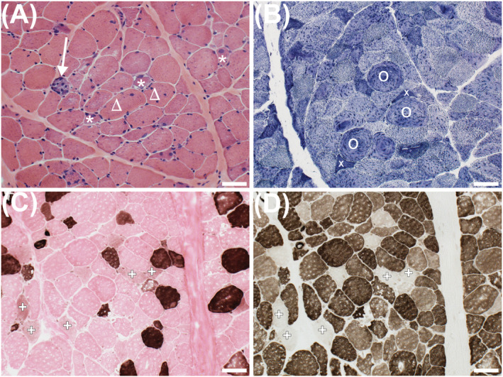
Figure 1.

Frozen sections of the vastus lateralis from a young female patient hospitalized with Covid‐19. Haematoxylin and eosin stain (A) shows a necrotic fibre replaced by macrophages (arrow), regenerating fibres (stars), one of which has subsarcolemmal vacuoles, and fibres with internalized nuclei (triangles). NADH‐reacted section (B) shows atrophic angulated fibres (x) and a disrupted mitochondrial network (O). ATPase‐reacted sections show type I fibres (dark at pH 4.3; C). At pH 9.4 (D), both type I and type II fibres are stained. Failed staining indicates the absence of functional myosin ATPase, indicative of myosinolysis (+; C, D). Scale bar = 50 μm.
