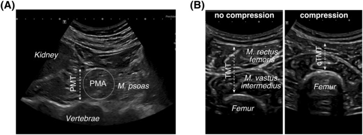
Figure 1.

Sonographic muscle quantification. Transversal sonographic B‐mode section (A) acquired at the height of the iliac cristae. The lower kidney pole, psoas muscle thickness (PMT), psoas muscle area (PMA) and vertebrae are indicated. Transversal sonographic B‐mode section (B) of the upper thigh middle, showing the thigh muscle group (Musculus vastus intermedius, Musculus rectus femoris) and the femur. Thigh muscle thickness without compression (TMT) and with compression (cTMT) by the sonography probe are indicated. All morphometric variables are obtained from a person in supine position.
