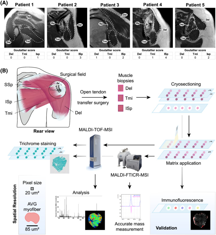
Figure 1.

Schematic overview of the study workflow. (A). Images of magnetic resonance imaging with arthrography of the shoulder muscles from the same patients whose muscle biopsies were used for MSI. The Del, Tmi and ISp regions are encircled with a dashed line. Goutallier score estimates fat accumulation in the respective muscles. (B). A rear schematic view of the diseased shoulder. Encircled is the surgical field with torn tendons that are located under the Del. Biopsies were taken from Del, Tmi and ISp close to the torn location during an open rotator cuff tendon transfer surgery. Sequential cryosections were scanned and analysed by both high‐spatial resolution (MALDI‐TOF) and high‐mass resolution (MALDI‐FTICR) MSI in negative and positive ion mode and subsequently were Gomori‐trichrome stained. Validation studies of MSI analyses were carried out on sequential sections.
