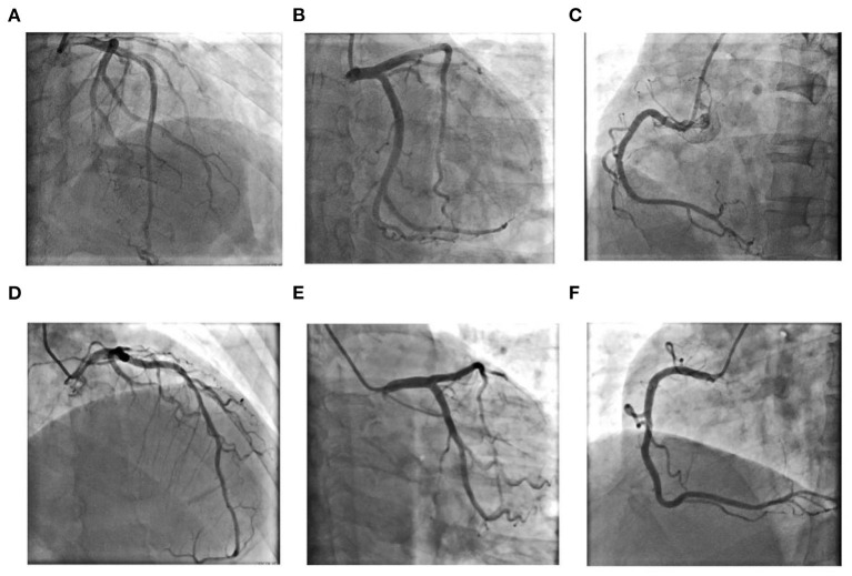
Figure 2.
Coronary angiography results for Case 1 (A–C): (A) Left coronary artery: Cranial 30°; (B) Left coronary artery: Caudal 30°; (C) Right coronary artery: Left anterior oblique 45°; Coronary angiography results for Case 2 (D–F): (D) Left anterior oblique 30°+ Cranial 30°; (E) Left coronary artery: Caudal 30°; (F) Right coronary artery: Left anterior oblique 45°.

