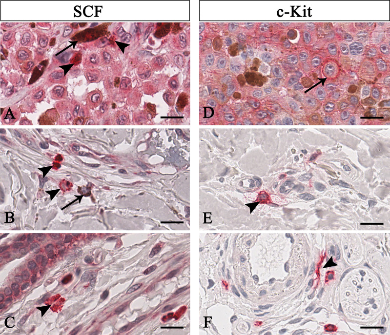
Figure 1.
Representative immunohistochemistry of SCF (A–C) or c-Kit (D–F) in the melanomas tissue sections. Micrographs show pigmented tumor cells (A, arrow) or not-pigmented tumor cells but with nucleolated nuclei (D, arrow) labelled for both SCF or c-Kit. Instead, SCF+/c-Kit+ mast cells are rounded-shape and hyperchromatic cells with positive granules in the cytoplasm (B, arrowhead) or secreted (A, C, E arrowhead). Some mast cells are spindle-shaped, thus resembling fibroblasts (F, arrowhead). Scale bar: 60 μm.

