Figure 2.
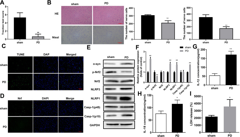
Oxidative stress and pyroptosis were promoted in the rat PD model. (A) Impaired muscle strength and equilibrium in rats of the PD model group. Motor dysfunctions in PD model rats were assessed by traction test scores. (B) Morphological changes in hippocampus were assessed by H&E and Nissl staining, In addition, Nissl-stained cells were evaluated in whole brain tissues of PD model rats. And the number of nerve cells was counted. (C) Increased numbers of apoptotic cells in brain tissues of the rat PD model group. TUNEL staining was used to detect cell apoptosis. (D) Changes in Nrf2 expression and subcellular locations in brain tissues of the rat PD model. Nrf2 expression and distribution were analyzed by immunofluorescence. (E) Protein abundances of PD and pyroptosis markers and phosphorylated Nrf2 in the rat PD model. Protein levels in rat tissues were quantified by Western blotting. (F) Relative protein levels of were quantified for westerns blot based on gray values. (G and H) Increased levels of IL-1β and IL-18 in serum from the PD model group. ELISA was used to determine IL-1β (F) and IL-18 (G) levels in rat serum. (I) Enhanced LDH release from rats in the PD model group. *P < 0.05; **P < 0.01.
Abbreviations: PD, Parkinson’s disease; α-Syn, α-synuclein; Nrf2, nuclear factor E2-related factor 2; NLRP1/3, Nod-like receptor protein 1/3; IL-1β/18, interleukin 1β/18; LDH, lactate dehydrogenase.

