Figure 5.
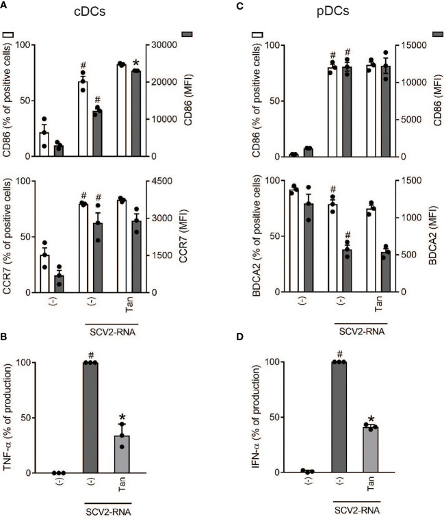
Effect of Tanimilast on primary DC activation by SCV2-RNA. cDCs (2x10⁶/ml) and pDCs (1x10⁶/ml) were pre-treated with Tanimilast (Tan, 10-7M) and then stimulated with SCV2-RNA for 24 hours. (A, C) The surface expression of CD86, CCR7 and BDCA2 was evaluated by FACS analysis. Data are expressed as the mean ± SEM (n=3) of the percentage of positive cells (left y axis) and of the Median Fluorescence Intensity (MFI) (right y axis). (B, D) The production of TNF-α and IFN-α was evaluated by ELISA in cell-free supernatants. Cytokine expression is normalized to SCV2-RNA condition (represented as 100%). Absolute levels of SCV2-RNA induced cytokines (ng/ml) were: TNF-α= 20.92 ± 0.55; IFN-α= 169.36 ± 23.39. Data are expressed as mean ± SEM (n=3). (A–D) #P< 0.05 versus (-) and *P< 0.05 versus SCV2-RNA by one-way ANOVA with Dunnett’s post-hoc test.

