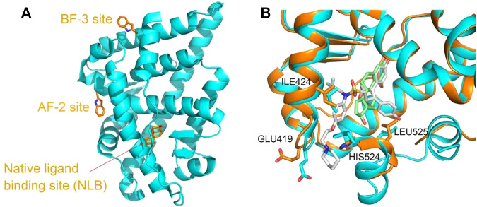
FIGURE 1.
(A) Three binding sites in the ligand-binding domain of the AR depicted based on PDBID 2POI and (B) the ligand-binding site flexibility of ERα in complex with different compounds, estradiol ((8R,9S,13S,14S,17S)-13-methyl-6,7,8,9,11,12,14,15,16,17-decahydrocy clopenta[a]henanthrene-3,17-diol) and 7AI ((1S,2R,4S)-5,6-bis(4-hydroxyphenyl)-N-{4-[2- (piperidin-1-yl)ethoxy]phenyl}-N-(2,2,2-trifluoroethyl)-7-oxabicyclo[2.2.1]hept-5-ene-2-sulfonamide) (structures from PDBID 5GS4 and 7RRX). Proteins are represented in a cartoon model, and compounds or residues, in a stick model. In (B), proteins from 5GS4 and 7RRX are colored in cyan and orange, and the compounds estradiol and 7AI are colored in green and gray, respectively. Residues undergoing considerable conformation changes, such as ILE 424, GLU419, HIS524, and LEU525 are depicted in the stick model.

