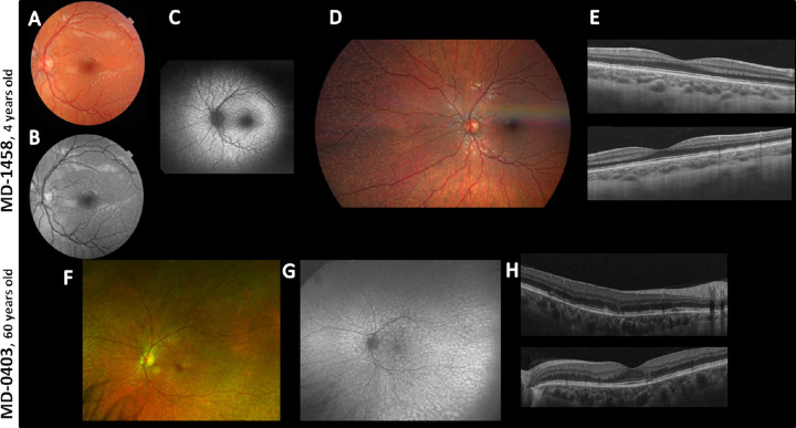
Figure 3.
Ophthalmological images of patients MD-1458 and MD-0403 with the novel variant p.(Cys103*) in homozygosis in the PLA2G5 gene. (A and F) Fundus images of both patients revealed flecklike lesions perifoveally within arcades with foveal sparing (similar in both eyes, only left eye shown). (B) Infrared reflectance image showed hyporeflective lesions perifoveally within arcades in both eyes (left eye shown) that correspond with hypopigmented lesions in the color picture in MD-1458. (C) Autofluorescence showed hyperautofluorescent scatter lesions that correspond with the hypopigmented lesions of the color picture. (D) Color fundus photograph of the right eye showing nasal periphery disclosing scatter yellowish dots outside the arcades. (E) OCT images showed RPE alterations (up, right eye; down, left eye). (G) Ultra-widefield fundus autofluorescence revealed hyperautofluorescent scatter lesions that correspond with the hypopigmented lesions of the color picture in both eyes (left eye shown). (H) OCT images showed RPE alterations in the macula (up, right eye; down, left eye).

