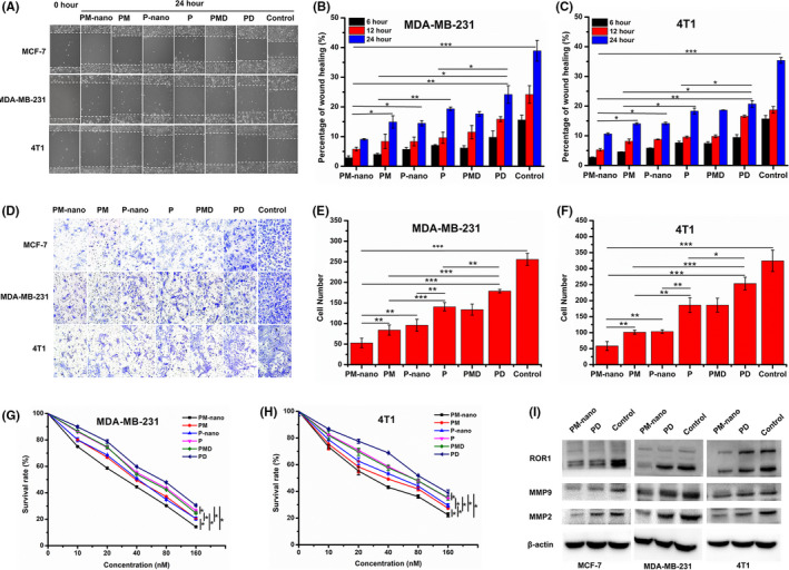
FIGURE 5.

Effects on the biological functions of breast cancer cells. A‐C, Images and quantification analysis of cell scratch assays before and after 24 h of treatment with various materials. D‐F, Images and quantification analysis of invaded cells in Transwell assays. G, H, Cytotoxicity of various materials against MDA‐MB‐231 cells and 4T1 cells evaluated by MTT assays. I, Western blots of receptor tyrosine kinase like orphan receptor 1 (ROR1), MMP9, and MMP2 in different cells after various treatments. *P < .05, **P < .01, ***P < .001. P, paclitaxel; PD, paclitaxel + dexamethasone; PM, paclitaxel + mifepristone; PMD, paclitaxel + mifepristone + dexamethasone; PM‐nano, paclitaxel‐conjugated and mifepristone‐loaded supramolecular hydrogel; P‐nano, paclitaxel hydrogel
