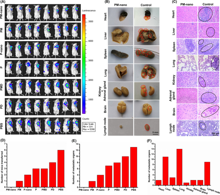
FIGURE 7.

Antimetastatic potential in vivo. A, Bioluminescence images of 4T1‐Luc tumor‐bearing mice on day 21 after treatment. B, C, Images of organs with and without breast cancer metastasis confirmed by gross observation (B) and H&E staining (C). The control group was defined as positive control and negative control groups, including paclitaxel + mifepristone (PM), paclitaxel hydrogel (P‐nano), paclitaxel (P), paclitaxel + mifepristone + dexamethasone (PMD), paclitaxel + dexamethasone (PD), and PBS groups. D, Number of mice with metastasis in each group evaluated by pathologic analysis. E, Number of organs with metastasis in each group confirmed by pathologic analysis. F, Number of organs with metastasis in all groups detected by pathologic analysis. PM‐nano, paclitaxel‐conjugated and mifepristone‐loaded supramolecular hydrogel
