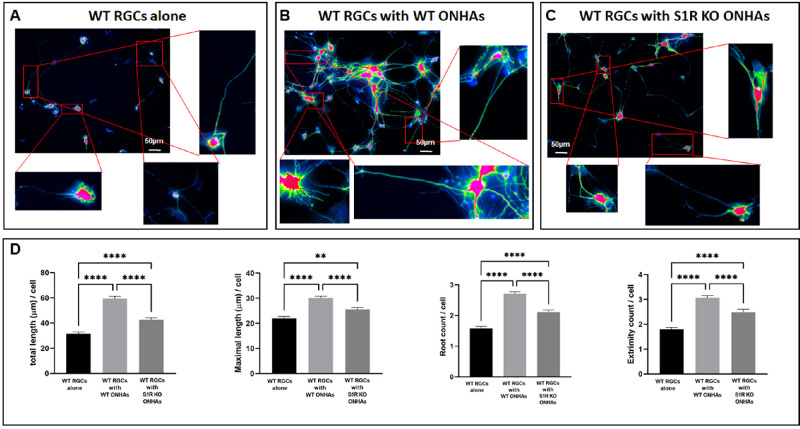
Figure 4.
RGC neurite outgrowth after 7 days. WT RGCs were isolated from neonatal mouse pups and cultured for 7 days. Scale bar, 50 µm. (A) WT RGCs alone were plated on the coverslips. (B) WT RGCs on coverslips were placed on the bottom of the lower chamber, and WT ONHAs were seeded on the membrane of the upper chamber. (C) WT RGCs on coverslips were placed on the bottom of the lower chamber, and S1R KO ONHAs were seeded on the membrane of the upper chamber. (D) RGC neurites were traced using Image J, Simple Neurite Tracer function, with green-colored overlay reflecting traced neurites. The total and maximal neurite length were measured and root and extremity numbers were counted. Significantly different from control *P < 0.05, **P < 0.01, ****P < 0.0001. Data were analyzed using one-way ANOVA followed by Tukey-Kramer post hoc test for multiple comparisons. Two coverslips, and eight microscopic fields per coverslip were quantified from each group of each isolation. The total number of cells analyzed per group was N = 120–380. These experiments were repeated in triplicate with cells isolated from different dates, different animals.

