Figure 1.
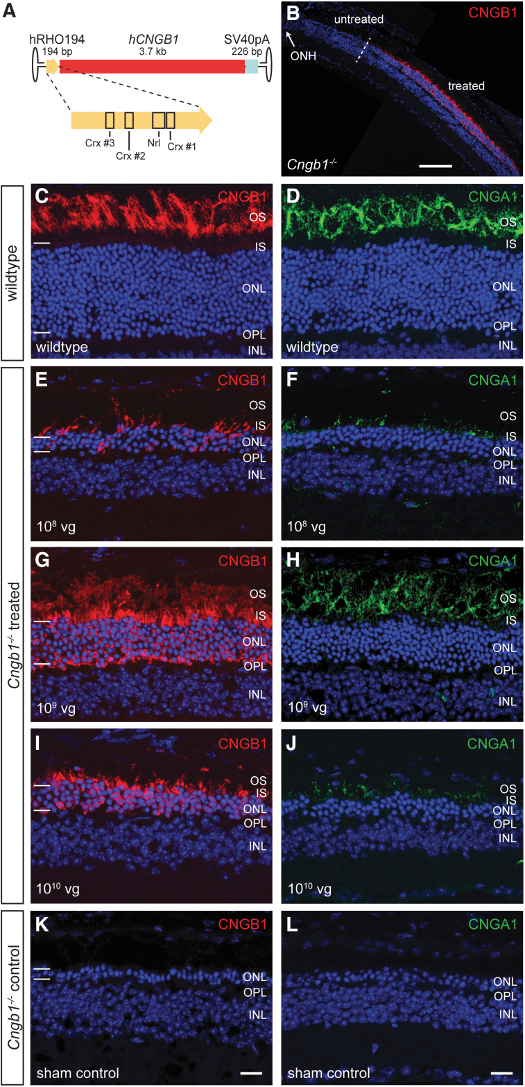
rAAV5.hCNGB1 results in a dose-dependent, efficient, rod-specific transgene expression in the Cngb1−/− retina. (A) Cartoon illustrating the gene expression cassette of rAAV5.hCNGB1. The novel human rhodopsin promoter is shown in higher detail and important transcription factor binding sites are marked. (B) Representative overview image of a confocal scan through a treated Cngb1−/− mouse retina covering treated (within the subretinal bleb) and untreated (outside the subretinal bleb) parts. The treatment border is marked with a dashed line. (C–L) Representative confocal images showing expression of CNGB1 (red, the antibody detects mouse and human CNGB1) and CNGA1 protein (green) in retinal cross sections of wild type (C, D), treated (E–J), and sham-injected control Cngb1−/− mouse retinae (K, L) at 10 months of age (9 months postinjection). Cngb1−/− mice were injected at 4 weeks of age with rAAV5.hCNGB1 at low dose (108 vg; E, F), mid-dose (109 vg; G, H) or high dose (1010 vg; I, J). Dependent on the dose, varying amounts of hCNGB1 protein (red) were found in the OS, the IS, and the ONL of all treated Cngb1−/− mice. Endogenous CNGA1 (green) expression was rescued in a dose-dependent manner and the protein localized to the OS of the treated area. Cell nuclei were stained with DAPI (blue). The edges of the ONL are marked with horizontal bars (C, E, G, I and K). GCL, ganglion cell layer; ONH, optic nerve head; OPL, outer plexiform layer. Scale bar in (B) marks 100 μm. Scale bar for (C–L) are shown in (K) and (L) and mark 25 μm. Cngb1, channel subunit beta 1; IS, inner segments; ONL, outer nuclear layer; OS, outer segments; vg, vector genomes. Color images are available online.

