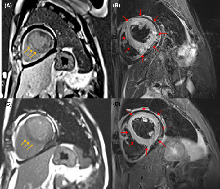
FIGURE 2.

Cardiac magnetic resonance (CMR) findings. (A) CMR imaging shows linear mid‐myocardial late gadolinium enhancement (LGE) of the septum wall at the base of the mid‐ventricle (yellow arrows). (B) T2‐weighted short‐axis inversion recovery imaging shows a high intensity of the global walls of the left ventricle suggestive of myocardial edema (red arrows). CMR findings met the original Lake Louise criteria for the diagnosis of acute myocarditis. On CMR imaging after 5 weeks, LGE disappeared (C), and the high intensity of the global walls of the left ventricle was sustained (D)
