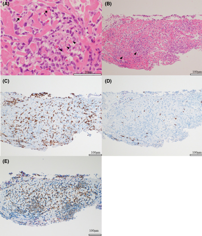
FIGURE 4.

Histopathological Findings. Endomyocardial biopsy of the right ventricular septum was performed. (A, B) Hematoxylin–eosin staining of heart tissue specimens obtained by endomyocardial biopsy shows myocarditis with inflammatory cells and cardiomyocyte damage (arrowheads). A few eosinophils are observed (arrows). (C, D) Immunostaining shows more infiltration of T lymphocytes of CD3‐positive cells than those of CD20‐positive cells. (E) Immunostaining for CD68 shows the infiltration of several macrophages
