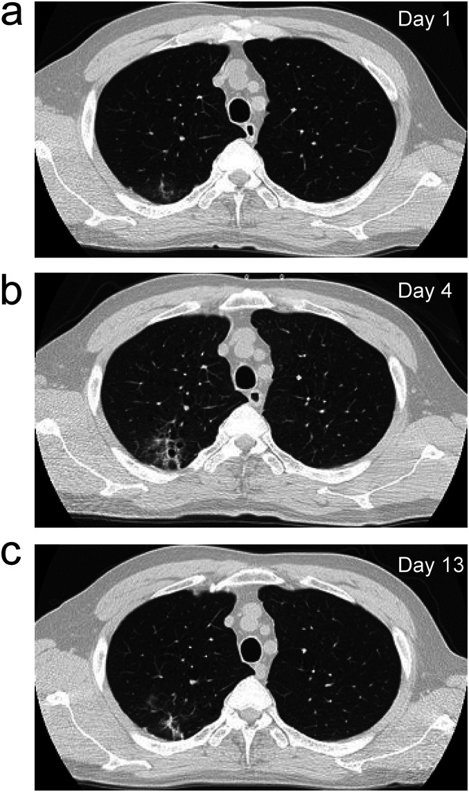
Figure 1.
Chest computed tomography images. (a) Image taken on Day 1 showing emphysematous changes in the lung field and ground-glass opacities with unclear borders below the dorsal pleura of the upper right lobe (b) Image taken on Day 7 showing slight exacerbation of the ground-glass opacities. (c) Image taken on Day 13 showing improvement in the ground-glass opacities.

