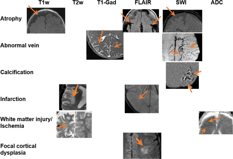
Figure 3.
Typical abnormalities (different rows) found in the brain MRI of patient with SWS, in multiple MRI sequences (different columns). Different figure panels are from different patients. Orange arrows point out the abnormal regions. ADC, apparent diffusion coefficient; FLAIR, fluid-attenuated inversion recovery; SWI, susceptibility-weighted imaging; SWS, Sturge-Weber syndrome.

