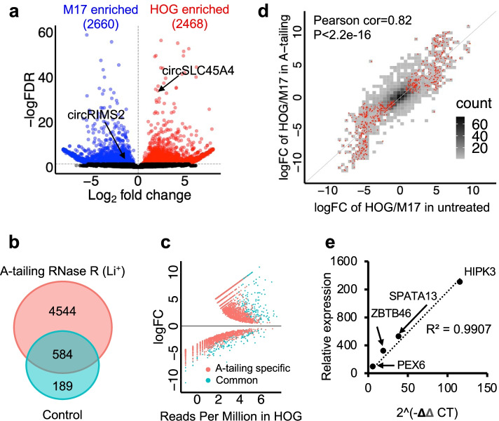
Fig. 2.
CARP identified a distinct circRNA landscape in M17 and HOG cells more efficiently using A-tailing data. a The volcano plot showed significant DE circRNAs in M17 and HOG cells using A-tailing data. Blue and red dots indicate significant M17 and HOG cell-enriched circRNAs (DESeq2, FDR < 0.05). b Overlap of DE circRNAs identified by CARP using A-tailing data and control data without A-tailing. c A scatter plot showing a log2 fold change of DE circRNAs in HOG and M17 cells and their expression (counts per million) in HOG cells. Red dots indicate DE circRNAs that were explicitly identified by A-tailing data. Cyan dots show DE circRNAs identified by both A-tailing data and control data. A-tailing libraries were sensitive in identifying circRNAs with relatively low expression levels (red dots). d The density plot showed a high correlation of log2 fold change for common circRNAs identified in the A-tailing library and the untreated library. Density colors show circRNA numbers in specific log2 fold changes. Red dots represent significantly DE circRNAs identified by A-tailing data (DESeq2, FDR < 0.05). e A scatter plot showed a high correlation of circRNA expression quantified by A-tailing data and qPCR for 4 randomly selected circRNAs with different expression levels

