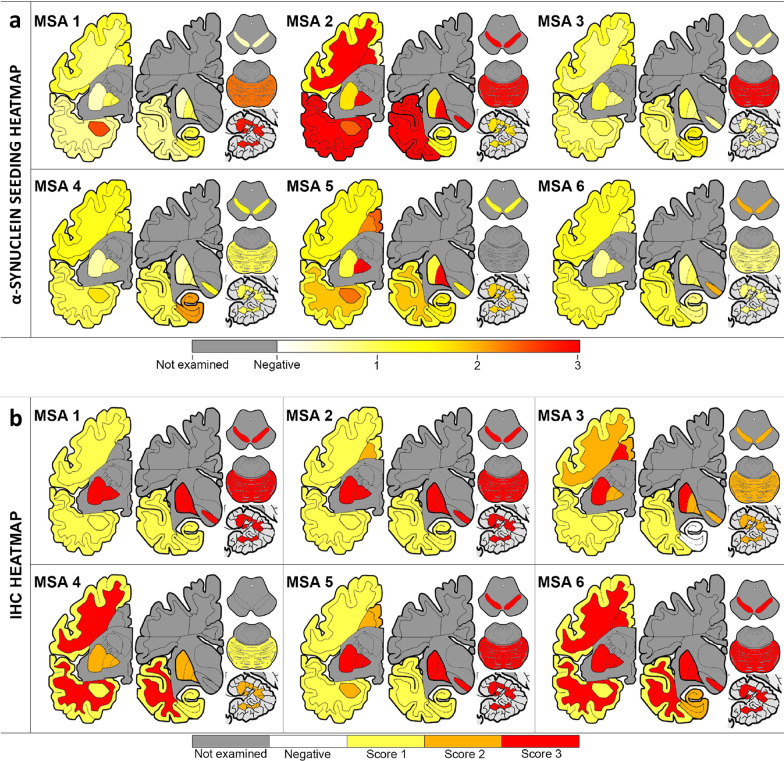
Fig. 6.
Extensive heterogeneity of α-synuclein seeding activity across different MSA patients and different brain regions. Heat mapping of α-synuclein seeding (a) assessed by RT-QuIC and of aggregated α-synuclein (b) evaluated by immunohistochemistry using the conformational α-synuclein 5G4 antibody. The α-synuclein seeding ranged from white (none) through yellow (low) and orange (medium) to red (high). The semiquantitative score of the severity of α-synuclein pathology ranged from white (none) through yellow (mild) and orange (moderate) to red (severe). Grey colored cortical regions indicate that the region was not evaluated

