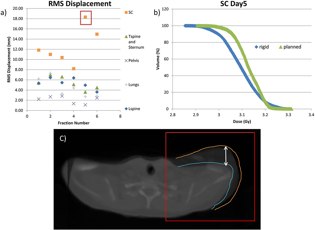
Fig. 5.
Example of misaligned shoulder from TMI treated patient. (a) root-mean-square (RMS) displacement per fraction showing a large shift in the SC region on days 5 and 6. (), and (b) dose-volume-histogram (DVH) graph of the rigid and planned dose of the shoulder, for day 5 of treatment (c) image of misaligned shoulder on day 5 of treatment.

