
FIGURE 2.
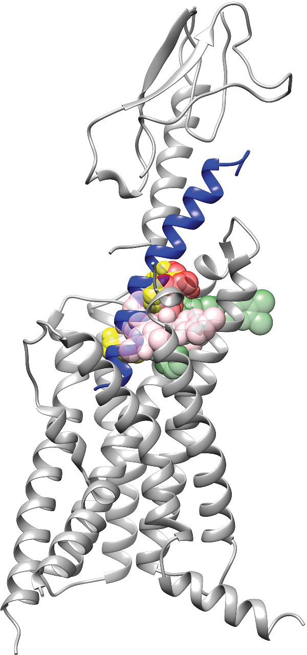
Binding sites of non‐peptide agonists of GLP‐1 receptors. The structure of the GLP‐1 receptor is shown (ECD: PDB ID: 6VCB; TMD, PDB ID: 6VCB), and the GLP‐1 peptide (PDB ID: 6VCB) is coloured in blue. Agonists are shown as coloured spheres. Dark green, TT‐OAD2 (PDB ID: 6ORV); yellow, RGT1383 (PDB ID: 7C2E); pink, LY3502970 (PDB ID: 6XOX); and red, PF06882961 (PDB ID: 6X1A). The opacity for the dark green and red colour was set at 47.5% and for the yellow and pink colour, 60%