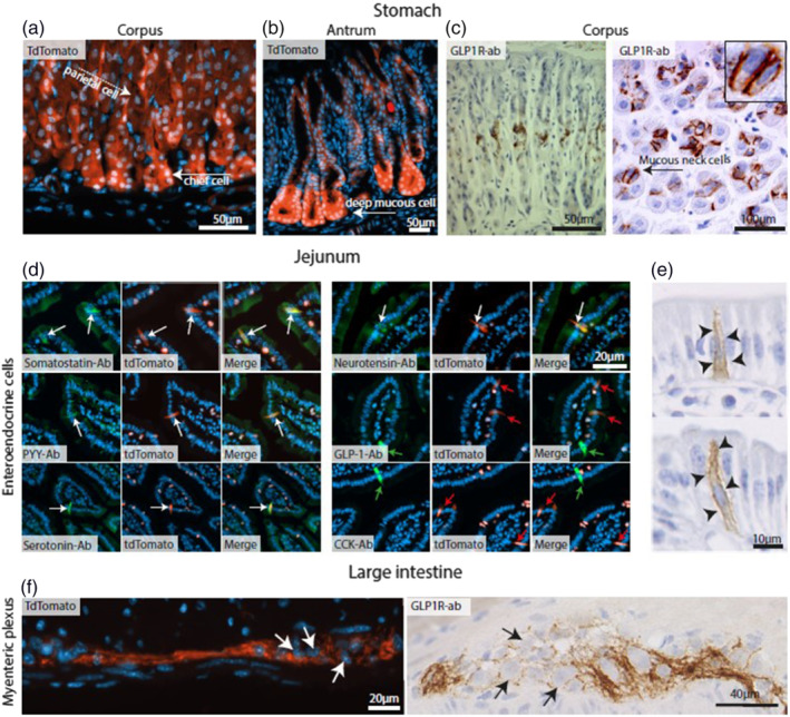
FIGURE 2.

Representative images of gastrointestinal cells expressing GLP‐1 receptors (GLP‐1R) cells. (a) Sections from mouse Glp1r.tdTomato gastric corpus showing Glp‐1r expression in chief cells (white arrow) and parietal cells (white dashed arrow). (b) Sections from mouse Glp1r.tdTomato gastric antrum showing Glp1r‐positive deep mucous cells (white arrow). (c) Sections of rat gastric corpus showing mucous neck cells (black arrow) stained with GLP‐1r antibodies (brown). (d) Glp1r.tdTomato expressing enteroendocrine cells (red cells) in the proximal small intestine co‐localizing (white arrows) with antibodies (green cells) specific for somatostatin, peptide‐YY, 5‐HT (serotonin), and neurotensin, but not GLP‐1 and CCK (green and red arrows) with merged pictures. (e) GLP‐1 receptor antibody staining (brown) in the rat proximal small intestine shows this receptor localizing to the lateral membrane of enteroendocrine cells (arrowheads). (f) Murine large intestine myenteric plexuses stained with Glp1r.tdTomato (red) or GLP‐1 receptor antibodies (brown). Note that expression of GLP‐1 receptors is exclusively found on fibres around the neurons, but not in their soma. Counterstaining performed with either DAPI or haematoxylin
