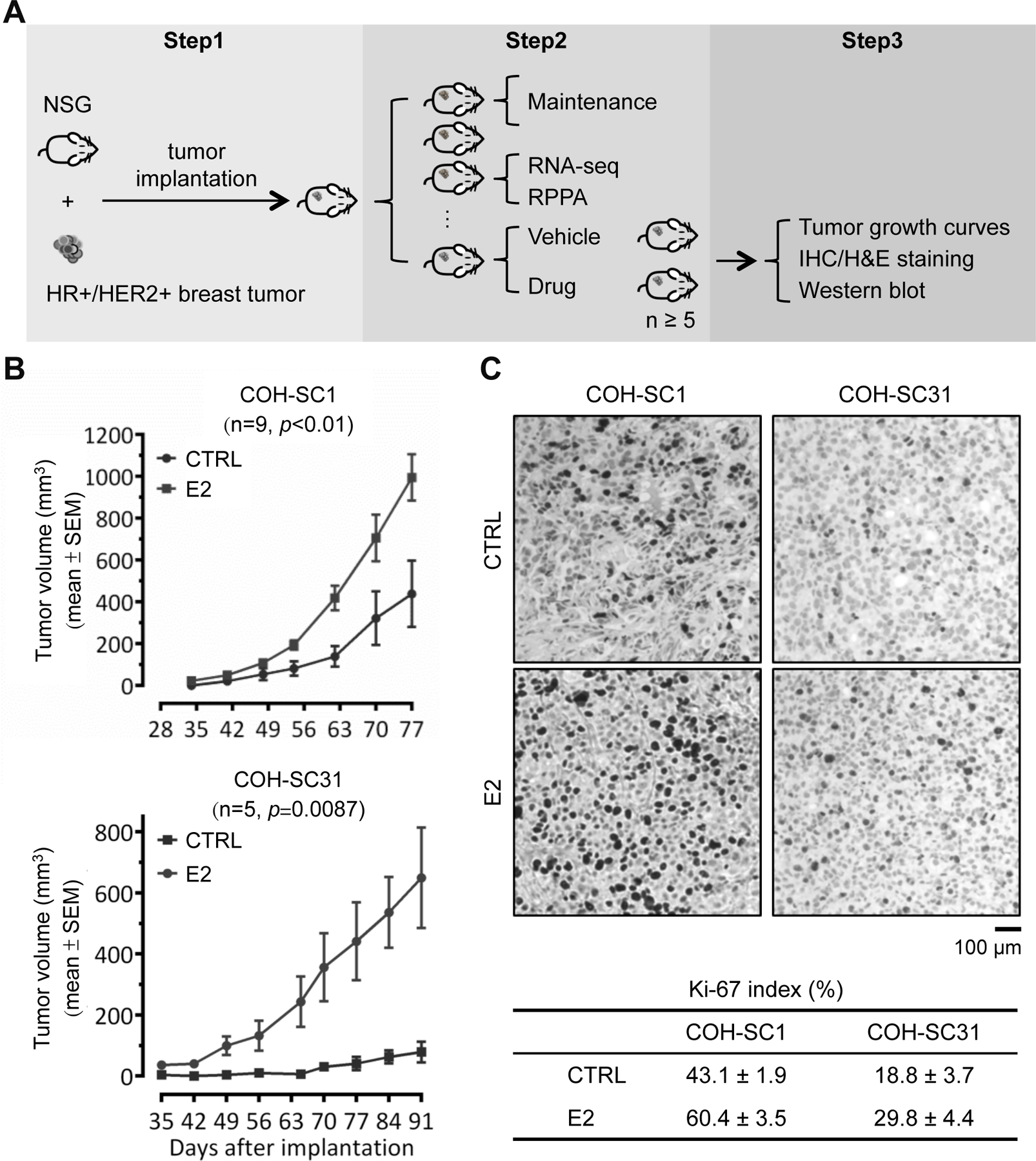
Figure 1. Differential estrogen response in COH-SC1 and COH-SC31 PDXs.

(A) Scheme of breast PDX establishment and utilization. Step 1, establishment of PDXs from breast cancer patients’ tumors; Step 2, stock expansion and molecular feature dissection of the PDXs; Step 3, in vivo drug efficacy examination and the subsequent histological and biochemical validation. (B) Estrogen-involved tumor growth in COH-SC1 and COH-SC31 PDXs. Ovariectomized mice were randomized and implanted COH-SC1 (n=9; upper panel) or COH-SC31 (n=5; lower panel) tumor accompanied with 1 mg estrogen (E2) or vehicle (CTRL) pellets. Tumor growth curves were monitored and plotted after implantation as indicated and p value was determined by two-way ANOVA analysis. (C) Ki-67 expression in COH-SC1 and COH-SC31 PDXs responded to estrogen treatment. Representative images of Ki-67 IHC in COH-SC1 and COH-SC31 PDXs upon E2 treatment were shown in upper panel and the associated scoring was summarized as Mean ± SEM in lower panel. Scale bar, 100 μm.
