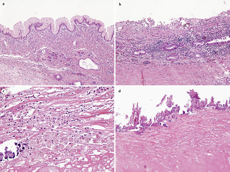
Fig. 2.
Representative microscopic images of commonly observed histologic features of pancreatic cyst after ablation therapy. a Residual mucinous lining epithelium with ovarian-type stroma (×200). b Moderate stromal lymphoplasmacytic infiltration (×200). c Foamy histiocytic aggregation with calcification (×400). d Diffuse calcification and stromal hyalinization along the cystic wall (×200).

