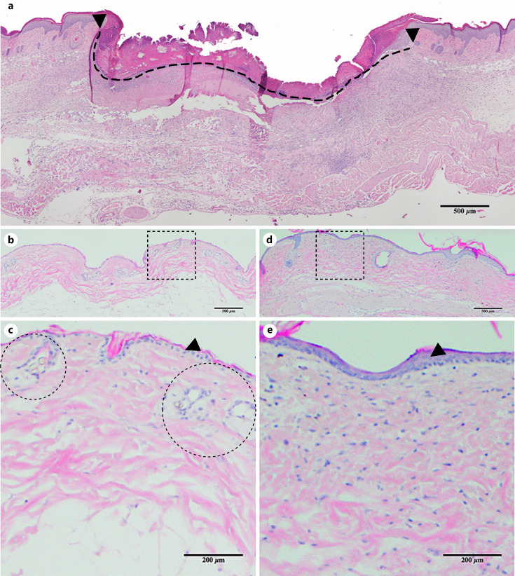
Fig. 1.
Wound cross-sectional histology and radiation changes in the skin. a Cross-section of the center of an untreated wound on POD12 showing the wound margin (black arrows) and wound surface diameter (dashed line, placed immediately below the scab). Mouse skin histology before (b), 31 days after (d) 40 Gy of radiation (i.e., POD21). c, e Magnified view of the dotted square above. Epidermal thickening (black arrows denote the epidermis) and increased collagen deposition is seen in the irradiated skin (d, e) compared to nonradiated skin (b, c). Note vessels within dermis (dotted circles in c) not seen in the irradiated skin (e). POD, postoperative day.

