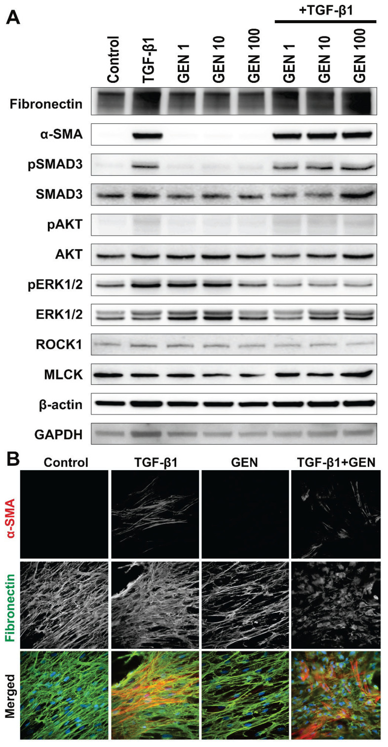
Fig. 1.
(A) Western blot analysis of human dermal fibroblasts exposed to genistein (GEN – 1, 10 and 100 nM) and/or TGF-β1 (30 ng/m); (B) Immunofluorescence analysis of human dermal fibroblasts (HDF) exposed to genistein (GEN – 100 nM) and/or TGF-β1 (30 ng/ml); α-SMA – alpha-smooth muscle actin (red signal); Fibr – fibronectin (green signal); nuclei were labeled with DAPI (blue signal) (magnification 600×).

