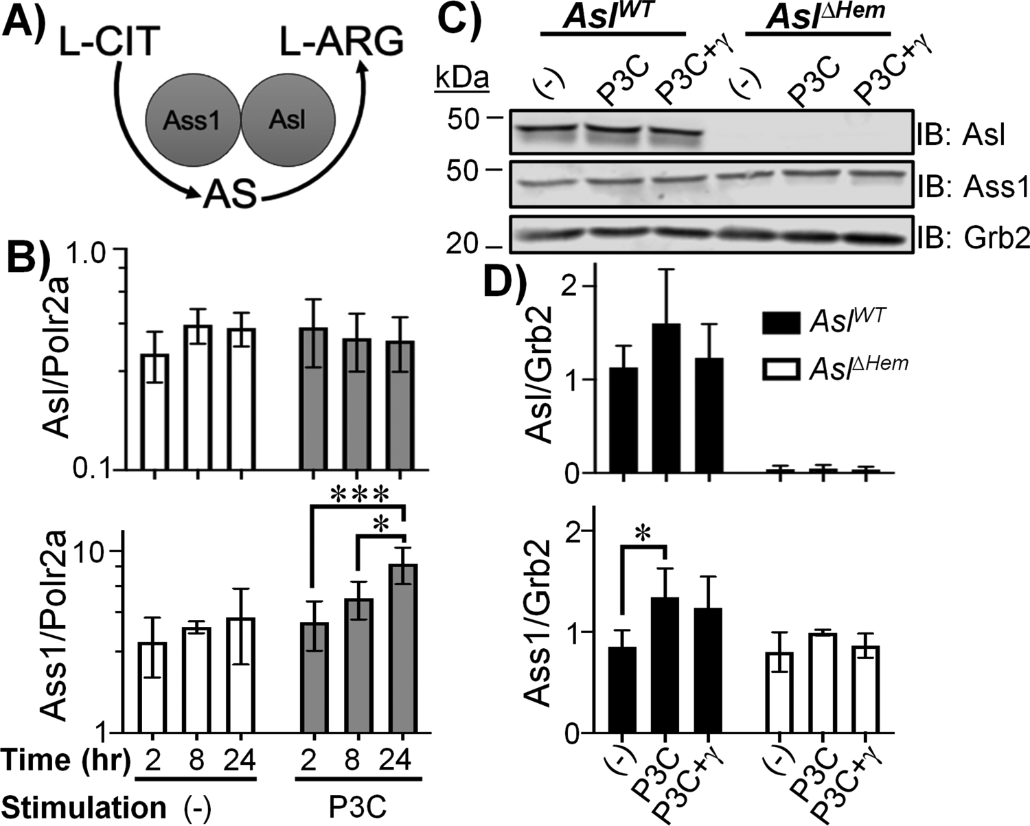
Figure 1. BMDCs express L-ARG synthesis enzymes.

(A) L-ARG synthesis pathway. (B) AslWT BMDCs were unstimulated (−) or stimulated with P3C for 2, 8, or 24h. Asl and Ass1 expression were determined by qRT-PCR. Data are mean±SD (N=6, 2 experiments). *p < 0.05, ***p < 0.001 by ANOVA with Tukey post hoc analysis. (C-D) AslWT and AslΔHem BMDCs were unstimulated (−) or stimulated with P3C±IFN-γ for 24h. Protein lysates were subjected to immunoblot (C) and densitometry analysis (D). Data are mean±SD (N≥3, 2 experiments). *p < 0.05, **p < 0.01 by Student’s t-test compared to (−) of same timepoint.
