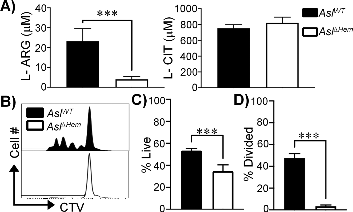
Figure 5. BMDC-synthesized L-ARG is found within the extracellular milieu and fuels CD4+ T cell viability and proliferation.

(A) L-ARG and L-CIT concentration in supernatants collected from co-cultures of AslΔHem CD4+ T cells with AslWT or AslΔHem BMDCs originally in media containing 1mM L-CIT. Data are mean+SD (N=5, 2 experiments). (B-D) CTV-stained AslΔHem CD4+ T cells were stimulated with α-CD3/CD28 and IL-2 for 96h. Media contained supernatants from AslΔHem CD4+ T cells co-cultured with AslWT (black) or AslΔHem (white) BMDCs originally supplemented 1mM L-CIT. (B) Representative live CD4+ T cell proliferation histograms. Graphical representation of live (C) and divided (D) cells. Data are mean+SD (N=6, 2 experiments). ***p < 0.001 by Student’s t-test.
