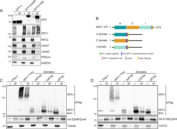
Fig 2. E248R and E199L interactions with endosomal proteins NPC1, Lamp 1 and Lamp2.
(A) GFP was immunoprecipitated in lysates from HEK293T cells transfected with EGFP, EGFP E248R or EGFP E199L. Representative immunoblot analysis of cell lysates (I) and GFP-immunoprecipitates (IP) using GFP, NPC1, NPC2, Lamp1, Lamp2 and PIKfyve antibodies. GAPDH was used as control. (n = 4 independent experiments, S2 and S3 Figs). (B) Schematic representation of NPC1 WT and specific recombinant constructions of individual domains shown in C and D. (C, D) HA was immunoprecipitated in lysates from HEK293T cells co-transfected with HA E248R (C) or HA E199L (D) with NPC1 Flag and FLAG NPC1 domains. Representative immunoblot analysis of total lysates (I) and HA-immunoprecipitates (IP) using Flag and HA antibody. Alpha-tubulin (C) or HSP90 (D) were used as control. “NPC1 dom.” refers to individual domains of NPC1 (n = 3 independent experiments, S5 and S6 Figs).

