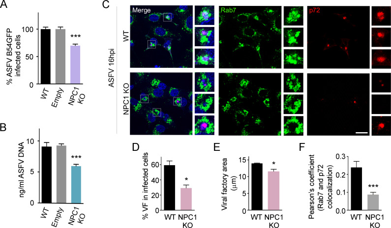
Fig 6. ASFV infection and replication in NPC1 KO cells.
Percentage of B54GFP infected cells (1 pfu/ml) at 16hpi in WT, Empty and NPC1 KO Vero cells detected by flow cytometry. Percentages were normalized to values in WT cells. (B) ASFV replication in WT, Empty and NPC1 KO ASFV infected cells quantified by real-time PCR. (C) Representative confocal images of WT and NPC1 KO cells stained for Rab7 (green), ASFV p72 (red) and DNA (Topro3, blue). Scale bar: 20 μm. Zoom images of ASFV viral factories (boxed regions) are also shown. (D) Percentages of viral factories in WT and NPC1 KO in infected cells shown in C. (E) Quantification of the cellular area occupied by viral factories stained with ASFV p72 in WT versus NPC1 KO infected cells shown in C. (F) Quantification of Rab7 and p72 colocalization in WT and NPC1 KO ASFV infected cells using ImageJ software. Graphs represent mean±sem from three independent experiments. Statistically significant differences are indicated by asterisks (***p < 0.001, **p < 0.01, *p < 0.05).

