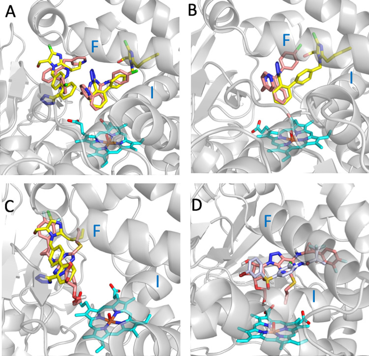
Fig 4. Docking conformations of vatalanib and ticagrelor in the binding pocket of CYP2C9.
(A) Two poses of vatalanib (in salmon) docked into the crystal structure of CYP2C9 (PDB ID 5XXI) and the two co-crystallized molecules of losartan (PDB ID 5XXI) (in yellow). (B) The best pose of vatalanib (in salmon) docked into the MD5 structure of CYP2C9 and the superposed co-crystallized structure of losartan of the PDB ID 5XXI (in yellow). (C) The best pose of ticagrelor (in salmon) docked into the crystal structure of CYP2C9 PDB ID 5XXI and one of the two co-crystallized molecules of losartan (PDB ID 5XXI) (in yellow). (D) The best pose of ticagrelor (in salmon) docked into the MD4 structure of CYP2C9 and the superposed co-crystallized structure of the CYP2C9 inhibitor 2QJ (PDB ID 4NZ2) (in gray). Helices F and I of CYP2C9 are noted. The MD4 and MD5 structures correspond to CYP2C9 conformations generated from MD simulations of CYP2C9 bound to losartan and apo CYP2C9, respectively.

