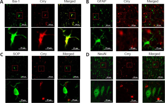
Figure 1.

Double immunofluorescence staining to identify the localization of Crry in the brains of 6-month-old P301S mice (original magnification 400×).
(A) Crry (FITC, green fluorescence) and the microglial marker Iba-1 (rhodamine, red fluorescence) colocalize in the brains. (B) Weak colocalization between Crry and the astrocyte marker GFAP (rhodamine, red fluorescence). (C) No colocalization between the oligodendrocyte marker OSP (rhodamine, red fluorescence) and Crry. (D) No colocalization between the neuron marker NeuN (rhodamine, red fluorescence) and Crry. Scale bars: 100 μm (upper) and 25 μm (lower). Crry: Cr1-related protein Y; FITC: fluorescein isothiocyanate; GFAP: glial fibrillary acidic protein; Iba-1: ionized calcium binding adaptor molecule 1; NeuN: neuronal nuclei; OSP: oligodendrocyte specific protein.
