Figure 6.
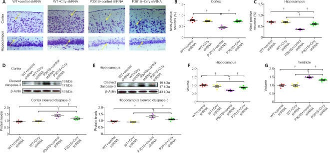
Decreasing Crry expression in the brain can delay neuron loss in P301S transgenic mice.
(A) Cresyl violet staining shows the survival of neurons in the cortex and hippocampus. Mice in the WT + control shRNA and WT + Crry shRNA groups have more neurons than P301S mice, and mice in the P301S + Crry shRNA group have more neurons compared with mice in the P301S + control shRNA group. Arrows indicate the neurons stained by cresyl violet. Scale bar: 100 μm. (B, C) Quantification of surviving neurons stained by cresyl violet in the cortex and hippocampus of mice in each group. (D, E) Cleaved caspase-3 expression in neurons of the cortex and hippocampus of mice in each group visualized by western blot. Protein expression levels were normalized to the WT + control shRNA group. (F, G) Quantitative analysis of hippocampal volume (F) and ventricle volume (G). Data are presented as the mean ± SEM (n = 6 for each group) and were analyzed by one-way analysis of variance followed by Tukey’s multiple comparison test. †P < 0.05. Crry: Cr1-related protein Y; shRNA: short hairpin RNA; WT: wild type.

