. 2022 Jan 4;11:e72195. doi: 10.7554/eLife.72195

Figure 3. Kruppel-like factor 9 (Klf9) functions as a brake on symmetric self-renewal of radial-glial neural stem cells (RGLs).

(A) Diagram of experimental design for in vivo two-photon imaging experiments. Inset is a high magnification image of a sparsely labeled single RGL in an adult Gli1CreERT2:Klf9+/+:Ai14 mouse. (B) Representative series of longitudinal imaging from four fields of view showing RGL symmetric and asymmetric divisions. Row 2: control. Rows 1, 3, and 4: experimental. Arrows point to mother cell and arrowheads point to daughter cells. Scale bar: 20 µm. (C) Quantification of RGL symmetric and asymmetric divisions showing an increase in symmetric divisions in Gli1CreERT2:Klf9f/f:Ai14 mice. n = 8 Gli1CreERT2:Klf9+/+:Ai14 mice, 65 divisions; n = 10 Gli1CreERT2:Klf9f/f:Ai14 mice, 77 divisions. Odds of symmetric division are 2.7× higher in Gli1CreERT2:Klf9f/f:Ai14 mice, p = 0.015 likelihood-ratio test., *(D) Similar number of divisions was recorded for each group to avoid biased assessment of division mode (n = 8 and 10 mice/group).

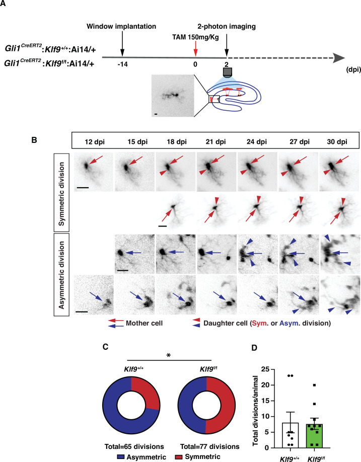
Figure 3.

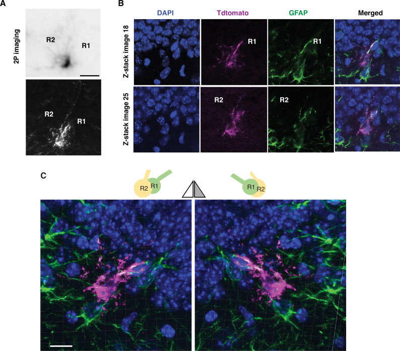
Figure 3—figure supplement 1. Representative images of radial-glial neural stem cell (RGL) divisions captured using two-photon imaging in vivo.

Figure 3—figure supplement 1.

(A) Representative two-photon images of RGL cells R1 and R2 in vivo and their respective post hoc fluorescence image. (B) Confocal immunofluorescence images of the same GFAP+/tdTomato+ cells at different depths, confirming their RGL identity. (C) Imaris deconvolution of tdTomato-labeled RGLs in B. Scale bar: 20 µm.
Figure 3—video 1. In vivo two-photon imaging of Gli1-postive radial-glial neural stem cells (RGLs).
Download video file (2.3MB, mp4)
Narrated example of longitudinal imaging of asymmetric neural stem cell (NSC) divisions. Two-photon imaging across days showing two examples of asymmetric division of NSCs (red arrows).
Figure 3—video 2. In vivo two-photon imaging of Gli1-postive radial-glial neural stem cells (RGLs).
Download video file (2.1MB, mp4)
Narrated example of longitudinal imaging of symmetric cell divisions. Two-photon imaging across days showing two examples of symmetric division of neural stem cells (NSCs; blue arrows).
Figure 3—video 3. In vivo two-photon imaging of Gli1-postive radial-glial neural stem cells (RGLs).
Download video file (28.5KB, mp4)
Three-dimensional reconstruction of RGL cells imaged in vivo before undergoing symmetric division. Field of view corresponds to second row of Figure 3B at 18 dpi.
Figure 3—video 4. In vivo two-photon imaging of Gli1-postive radial-glial neural stem cells (RGLs).
Download video file (30.5KB, mp4)
Three-dimensional reconstruction of RGL cells imaged in vivo after undergoing symmetric division. Field of view corresponds to second row of Figure 3B at 30 dpi.