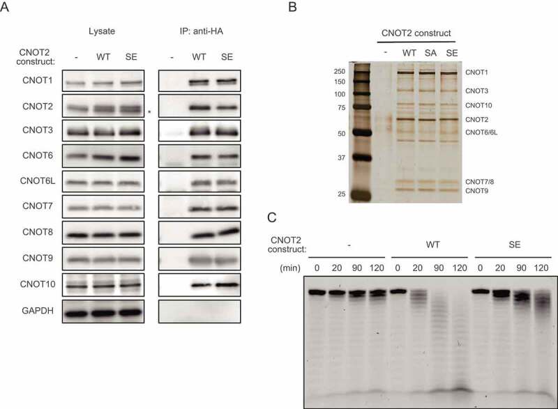
Figure 5.

(A) Lysates were prepared from HeLa cells expressing HA-CNOT2 WT (WT) or HA-CNOT2 SE (SE) and immunoprecipitated using anti-HA antibody. The anti-HA immunoprecipitates (IP) and cell lysates were analysed by immunoblot. HeLa cells infected with control retrovirus (-) were used as controls. An asterisk indicates endogenous CNOT2. Please see Supplementary Fig. S6B for results of HA-CNOT2 SA. (B) Lysates were prepared from control HeLa cells (-) and HeLa cells expressing HA-CNOT2 (WT, SA or SE) and immunoprecipitated using anti-HA antibody. The anti-HA immunoprecipitates were analysed by silver staining. CNOT complex subunits predicted from molecular masses (but not identified by mass spectrometry) are indicated. (C) IPs prepared as in (A) were incubated with 5ʹ-labelled poly(A) RNA for the indicated times. Reaction products were then analysed on a denaturing gel.
