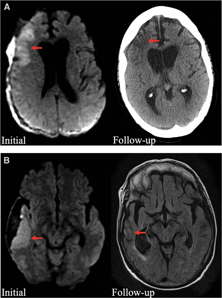
FIG. 3.
Representative long-term follow-up images for 2 patients with peri-SDH cytotoxic edema. (A) Initial diffusion-weighted MRI image shows cytotoxic edema in the right frontal lobe (left panel, red arrow), and follow-up CT at 2 months shows persistent encephalomalacia (right panel, red arrow). (B) Initial diffusion-weighted MRI shows cytotoxic edema in the right lateral temporal lobe (left panel, red arrow), and follow-up MRI at 1 year shows encephalomalacia and focal atrophy (right panel, red arrow). CT, computed tomography; MRI, magnetic resonance imaging, SDH, subdural hematoma.

