
Fig. 4.
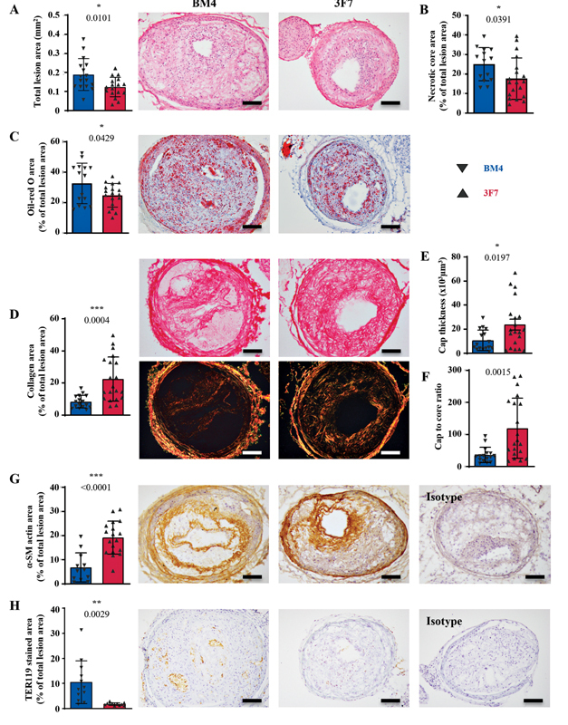
Inhibition of FXIIa via antibody 3F7 stabilizes vulnerable atherosclerotic plaques. ApoE −/− mice were placed on an HFD for 6 weeks prior to TS surgery. Following surgery, animals remained on the HFD for 7 weeks and 3F7 or BM4 isotype control was administered. ( A ) H&E was used to morphologically assess plaque size ( p = 0.0101; BM4 n = 16; 3F7 n = 17) and ( B ) necrotic core area ( p = 0.0391; BM4 n = 14; 3F7 n = 19) in unstable plaques, as compared with the BM4-treated animals. ( C ) Oil-red O staining was used to assess the lipid deposition within the plaques ( p = 0.0429; BM4 n = 15; 3F7 n = 18). Picro-Sirius Red was used to assess the degree of protective collagen deposition within the plaques, with histological examination performed using BF and polarized light. ( D ) The total intimal collagen deposition, ( E ) fibrous cap thickness, and ( F ) cap-to-core ratio, as compared with BM4-treated animals ( p -values = 0.0004; 0.0197; 0.0015; BM4 n = 16; 3F7 n = 20). ( G ) Intimal smooth-muscle cell deposition ( p = < 0.0001; BM4 n = 12; 3F7 n = 18) and ( H ) intra-plaque hemorrhage, as indicated by erythrocyte/TER119 staining ( p = 0.0029; BM4 n = 12; 3F7 n = 11). Isotype refers to the isotype control for the antibody used in the respective immunohistochemical stain. Assays were assessed using unpaired Student's t -tests. Values are mean ± SD. Scale bars = 200 µm. α-SM, α-smooth muscle; BF, brightfield; FXII, factor 12; FXIIa, activated factor 12; H&E, hematoxylin and eosin; HFD, high-fat diet; SD, standard deviation.