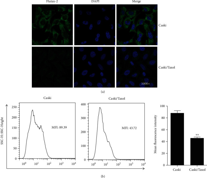
Figure 3.

(a) Intracellular green fluorescence was monitored by confocal microscopy, and (b) the strength of fluorescence was measured by flow cytometry. The results showed that fluorescence in the Caski/Taxol cells was significantly weaker compared with that in the Caski cells, indicating significantly lower intracellular drug concentrations of paclitaxel in the Caski/Taxol cells. ∗∗P < 0.01. Caski/Taxol: paclitaxel-resistant Caski cells.
