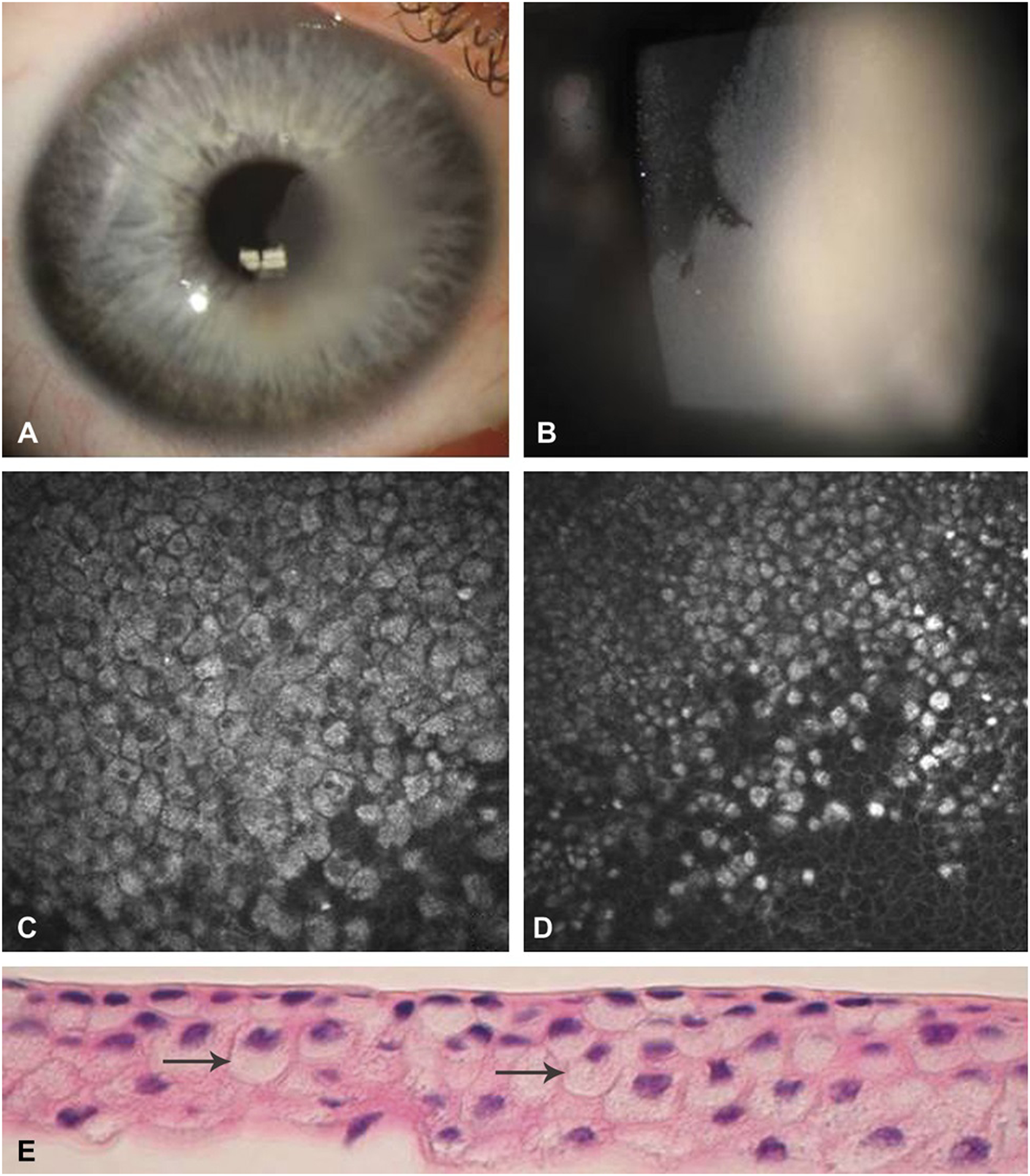
FIGURE 1.

Clinical presentation of Case 1. A, Slit-lamp photograph showing localized corneal epithelial opacification involving the visual axis. B, Higher magnification slit-lamp photograph of the corneal lesion in sharp contrast to adjacent normal epithelium. C-D, In vivo confocal microscopy of epithelial cells with hyperreflective cytoplasm and hyporeflective nuclei. E, Histopathologic demonstration of vacuolization in the cytoplasm of the involved epithelial cells (arrows).
