
FIGURE 3.
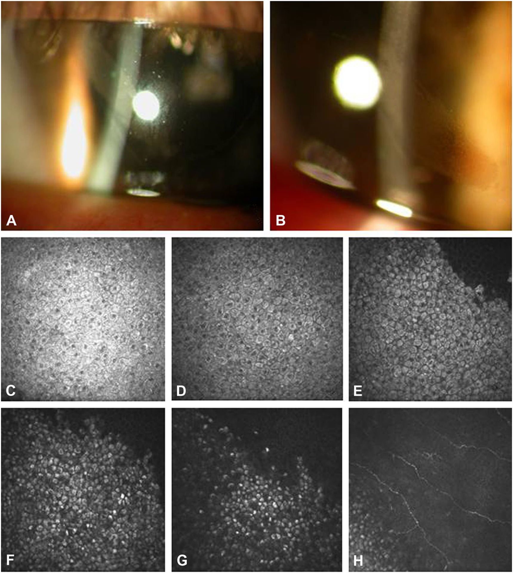
Clinical Presentation of Case 2. A, Slit-lamp photograph showing a band shaped corneal epithelial lesion radiating from the limbus. B, Higher magnification slit-lamp photograph showing microcystic pattern corneal opacification. C-G, In-vivo confocal microscopy from superficial to the wing epithelial layers (C-E) and basal epithelial layers (F-H) demonstrating cells with hyperreflective cytoplasm and hyporeflective nuclei. The nuclei are less prominent in the basal cells (F-G).