
FIGURE 4.
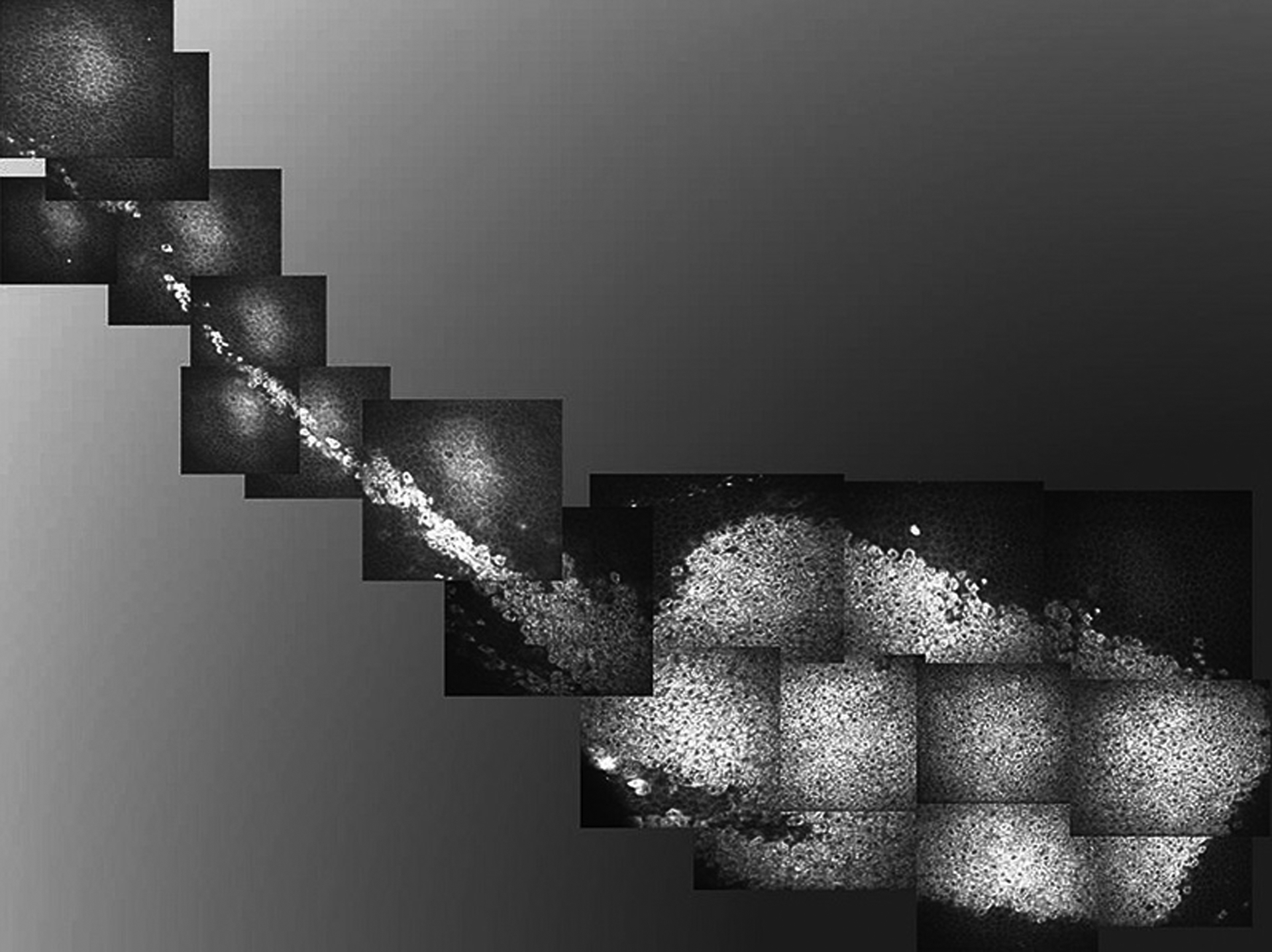
Case 2. In vivo confocal microscopy composite image demonstrating localized, well-demarcated distribution of affected epithelial cells with hyperreflective cytoplasm. The configuration of the confocal image corresponds to the clinical appearance and location of the epithelial opacification. The tapered appearance of the lesion is near the limbus (pictured at the left side of the image). The basal limbal epithelial cells were also involved.