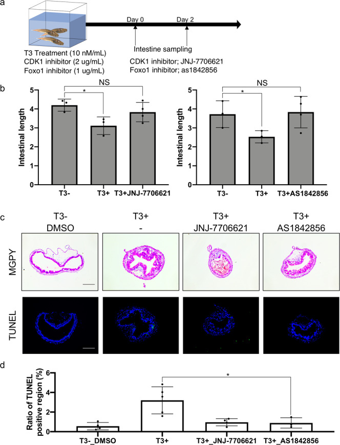
Fig. 7. Inhibiting CDK1 signaling blocks intestinal remodeling during T3-induced metamorphosis.

a Experimental design for treatment with CDK1 and Foxo1 inhibitors. Wild-type tadpoles at stage 54 were treated with 2 μg/ml of CDK1 inhibitor JNJ-7706621 or 1 μg/ml of phosphorylation inhibitor of Foxo1 as1842856 for 2 days in the presence and absence of T3. Most of tadpoles treated with the inhibitors in the presence of T3 died at 3 days. Thus, intestinal remodeling was analyzed after 2 days of T3 treatment. b JNJ-7706621 and as1842856 inhibits T3-induced shortening of the intestine. Tadpoles at stage 54 were treated with or without 10 nM T3 for 2 days in the presence or absence of indicated inhibitor. Intestinal length was measured from bile duct junction to colon and normalized against body length. Each group included more than 4 tadpoles. Each bar represents the mean plus S.E. The asterisk (*) indicates a significant difference between the T3 treated tadpoles and control animals (P < 0.05) (see Supplementary Data 21 for raw data). c T3-induced intestinal morphological changes (epithelial folding) and apoptosis in the intestinal epithelium were inhibited by JNJ-7706621 and as1842856. Tadpoles were treated as above. Cross-sections of the intestine were stained with MGPY, which stains DNA blue and RNA red (top), and apoptotic cells were detected by TUNEL staining (bottom). TUNEL positive cells were stained green and nuclei were stained by Hoechst 33342 (blue). Bar indicates 20 μm (see Supplementary Data 21 for raw data). d Quantification of the apoptotic cells in c. TUNEL positive epithelium region was measured by using ImageJ software and normalized against the total epithelium region as determined by DAPI staining (see Supplementary Data 22 for raw data).
