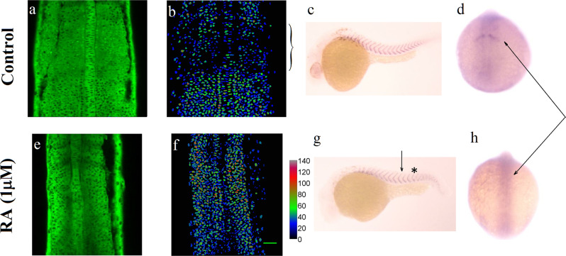
Fig. 3. Somitogenesis at high Erk activity.
a–d Control: normal development. a Erk activity in DREKA embryos at 14 somite stage: dark nuclei point to high Erk activity. b The false color images represent the Erk activity averaged over a 30 μm perpendicular stack of images. c ISH against xirp2a (a marker of somite boundaries) in 30 hpf embryos. d IHC against Mesp2a, a marker of the last somite boundary at 14 somites (arrow). e–h Somitogenetic development in DREKA embryos incubated from one-cell stage in 10 μM DEAB in presence of 1 μM RA from the ten somite stage (arrow in g). e, f The Erk activity is high throughout the PSM. g ISH against xirp2a in 30hpf embryos. Somitogenesis is impaired past 13 somites (indicated by a *) with unclear somite boundaries. h IHC against Mesp2a show that it is not expressed past 13 somite stage (arrows in d and h). Scale bar: 50 μm.

