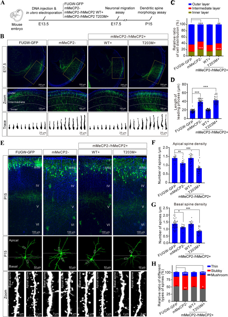
Fig. 5.
T203 O-GlcNAcylation is required for dendritic spine morphogenesis in vivo. A Schematic of the experimental design. B Upper panels, distribution of GFP+ pyramidal neurons in the indicated plasmid-electroporated neocortex at E17.5. Middle panels (zoom), higher magnification to illustrate the detailed distribution within the neocortex, showing the inner, intermediate, and outer layers. Lower panels (trace), Fiji tracings of representative single GFP+ neurons from the indicated groups. Scale bars, 200 μm, 100 μm, and 25 μm. C Relative ratios of GFP+ neuron distribution (%) in distinct neocortical layers (mean ± SEM, χ2-test, **P < 0.01, ***P < 0.001). D Length of the leading process (LP) in GFP+ neurons electroporated with the indicated plasmid (mean ± SEM, one-way ANOVA followed by the Bonferroni test, ***P < 0.001). E Representative images of dendritic spines on apical and basal dendrites of GFP+ neurons at P15 after electroporation with the indicated plasmid (Zoom panels, higher magnification to illustrate the detailed dendritic spine morphology; scale bars, 100 μm, 50 m, and 3 μm). F, G Spine density on apical and basal dendrites in GFP+ neuron (mean ± SEM, one-way ANOVA followed by the Bonferroni test, *P < 0.05, **P < 0.01, ***P < 0.001). H Distribution of three subtypes of dendritic spine in GFP+ neurons at P15 (mean ± SEM, χ2-test, *P < 0.05, **P < 0.01, ***P < 0.001).

