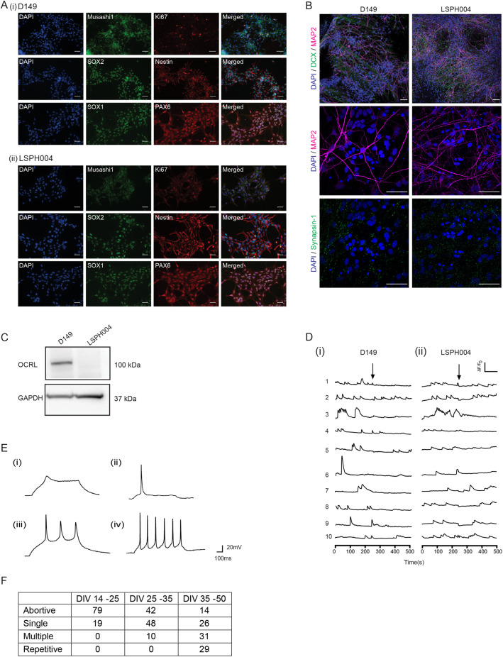
Fig. 2.
Characterization of NSCs and neurons derived from LS hiPSC. (A) Immunocytochemistry of NSCs (i) D149 (control) (ii) LSPH004 (patient) showing the expression of the NSC markers SOX1, SOX2, PAX-6, Nestin, Mushashi-1 and the proliferation marker Ki-67. Nucleus stained with DAPI. Scale bars: 50 μm. (B) Immunofluorescence images (maximum intensity projections) of D149 and LSPH004 neurons at 30 DIV differentiated from respective NSCs. The cells were stained with the following neuronal markers: DCX (green, immature neuronal marker) and MAP2 (magenta, mature neuronal marker); synapsin-1 (green) followed by counterstaining with DAPI (blue). Scale bars: 50 μm. (C) Western blot showing expression of OCRL protein in lysates from 30 DIV Neurons in the control line D149 and its absence in patient line LSPH004. GAPDH was used as a loading control. (D) Calcium transients recorded from 30 DIV D149 (i) and LSPH004 (ii) neurons are shown. Each panel shows [Ca2+]i traces from individual cells in the dish. Y-axis shows normalized fluorescence intensity ΔF/F0 and X-axis is time in seconds. The baseline recording for 4 mins, followed by addition of 10 μM tetrodotoxin (TTX) (as indicated by the arrows). (E) Evoked AP in neurons differentiated from NSC recorded using whole-cell patch clamp electrophysiology at 10, 20, 30 and 40 DIV. The characteristic feature of action potentials recorded from control D149 cells at each time point is shown. (Ei) Immature AP at 10 DIV, single action potential at 20 DIV (Eii) and (Eiii) multiple AP on 30 DIV (Eiv). Most neurons exhibited repetitive firing by 40 DIV. (F) Table depicting the proportion of neurons that showed abortive, single, repetitive and multiple action potentials as a function of age. Columns depict the age of the neurons as DIV. Rows depict the type of action potential. Each number shown is the percentage of cells recorded at that age, which showed a specific type of action potential.

