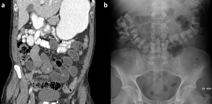
Figure 12.
Example of successful CT-based WSC challenge. (a) Initial CT which demonstrates dilated fluid-filled distal small bowel. Oral contrast was not near the transition point. (b) Follow-up AXR demonstrating transit of CT oral contrast into colon. AXR, abdominal radiographs; WSC, water-soluble contrast.

