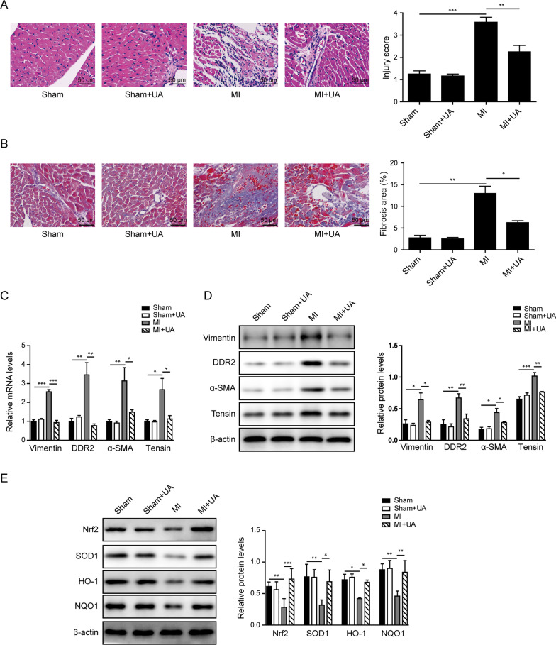
Fig. 5.
UA treatment inhibited myocardial fibrosis via Nrf2 pathway in vivo. A Represent images by H&E staining of heart tissues from indicated groups and statistic score of injury level. B Represent images by Masson’s trichrome staining of heart tissues from indicated groups and statistic results of the fibrosis area. C The mRNA levels and D protein levels of fibrosis markers including vimentin, DDR2, tensin and α-SMA in heart tissues from indicated groups. E The protein levels of Nrf2, SOD1, HO-1 and NQO1 in heart tissues from indicated groups. β-actin was used as normalized control. *P < 0.05, **P < 0.01, ***P < 0.001

