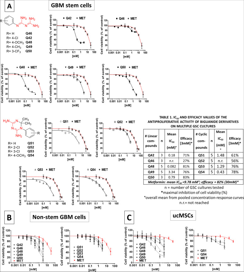
Fig. 1.
A Concentration-response curves of novel biguanide derivatives on GSC viability, in comparison with metformin activity. UPPER LINES (from the left): Chemical structures of linear biguanides (structurally related to metformin) highlighting the biguanide moiety (in red); antiproliferative activity of Q42, Q46, Q48, Q49, and Q50 in comparison with metformin activity. LOWER LINES (from the left): Chemical structures of cyclic biguanides (structurally related to cycloguanil) highlighting the biguanide moiety (in red); antiproliferative activity of Q51, Q52, Q53, and Q54 in comparison with metformin activity. Cell viability was evaluated by MTT assay after 48 h of treatment. Data are reported as average of replica experiments in multiple GSC cultures (mean ± S.E.M. of at least three independent experiments for each culture). Table 1 within the figure reports the number of cultures analyzed for each compound and the calculated potency and efficacy. Compound effect in individual cultures is reported in Fig. S3, and point-by point statistical analysis is reported in Table S3. B Concentration-response curves of novel biguanide derivatives on differentiated (non-GSCs) glioblastoma cell viability, in comparison with metformin activity. Left graph: antiproliferative activity of Q42, Q46, Q48, Q49, and Q50 in comparison with metformin activity (in red). Right graph: antiproliferative activity of Q51, Q52, Q53, and Q54 in comparison with metformin activity (in red). Cell viability was evaluated by MTT assay after 48 h of treatment. Data are reported as average of replica experiments in multiple differentiated glioblastoma cell cultures (mean ± S.E.M. of at least three independent experiments for each culture), obtained by the same GSC cultures analyzed in A, by shifting culture conditions in FBS containing medium. Compound effect in individual cultures is reported in in Fig. S4, and point-by point statistical analysis is reported in Table S4. C Concentration-response curves of novel biguanide derivatives on umbilical cord mesenchymal stem cell (ucMSC) viability, in comparison with metformin activity. Left graph: antiproliferative activity of Q42, Q46, Q48, Q49, and Q50 in comparison with metformin activity (in red). Right graph: antiproliferative activity of Q51, Q52, Q53, and Q54 in comparison with metformin activity (in red). Cell viability was evaluated by MTT assay after 48 h of treatment. Data are reported as average of replica experiments in independently isolated ucMSC cultures (mean ± S.E.M. of at least three independent experiments for each culture). Compound effect in individual cultures is reported in in Fig. S5, and point-by point statistical analysis is reported in Table S5

