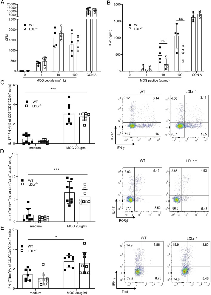
Fig. 2.
LDLr deficiency does not influence proliferation nor cytokine production induced by a T cell recall response. A On day 8 after immunization, splenocytes were isolated from WT and LDLr−/− mice and restimulated with MOG35-55 in vitro. The proliferative response was measured by [3H] thymidine incorporation 72 h after restimulation with different concentrations of MOG35-55 peptide or Concanavalin A (CON A) and expressed in counts per minute (CPM) (mean ± SD, n = 4 mice). B Cytokine IL-2 production in culture supernatants after 48 h of culture with the indicated concentration of MOG35–55 or CON A at 10 µg/ml was determined by ELISA (mean ± SD, n = 4 mice). C–E Flow cytometric analysis of the percentage of IL-17+IFN-γ+,IL-17+RORγt+ and IFN-γ+Tbet+ in CD3+CD4+CD44+ T cell at day 6 after restimulation of the indicated concentration of MOG35–55. Representative dot plots for C–E are results from WT and LDLr−/− splenocytes restimulated with 20 μg/ml of MOG35–55. Shown results are from mice pooled from two independent experiments (mean ± SD, n = 8 mice). Data are representative of three experiments. ∗p < 0.05, ∗∗∗p < 0.001, NS, not significant; p values were determined by two-way ANOVA with Sidak’s post hoc test (A–E)

